
Statin muscle pain: mechanism, CYP interactions and counselling points 2026
Myopathy mechanism, CYP interactions that raise risk, and counselling points. Safety-first pharmacology in six minutes.
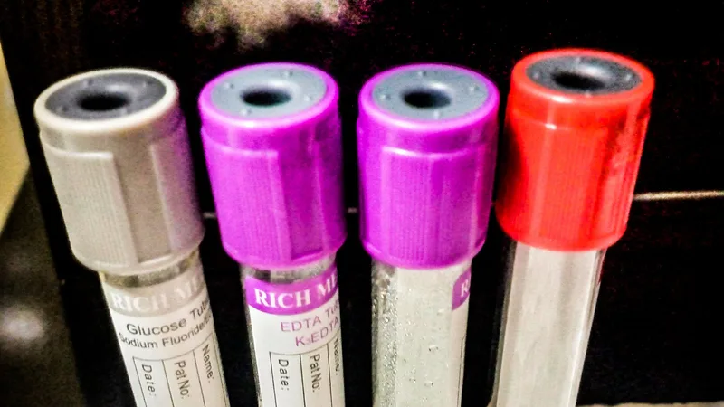
 Photo: Robert Flogaus-Faust / CC BY 4.0, via Wikimedia Commons
Short answer: digoxin inhibits the Na+/K+ ATPase pump, raising intracellular calcium. Excess inhibition produces arrhythmias from combined automaticity and conduction block. Hypokalaemia potentiates toxicity. Digoxin immune fab reverses life-threatening poisoning.
Cardiac glycosides derived from Digitalis purpurea (foxglove) inhibit the membrane Na+/K+ ATPase. Intracellular sodium concentration rises, reducing the sodium gradient that drives the Na+/Ca2+ exchanger. Intracellular calcium rises, increasing contractile force (positive inotropy). Vagal effects slow AV nodal conduction, providing rate control in atrial fibrillation. The therapeutic range is narrow: 0.5 to 2.0 ng/mL in most references, with toxicity possible even within range when potentiating factors coexist.
Excessive pump inhibition raises intracellular calcium beyond the therapeutic window. Calcium overload produces delayed afterdepolarisations, generating ectopic beats and tachyarrhythmias. Simultaneously, enhanced vagal tone and direct AV nodal effects slow conduction, producing bradycardia and AV block. The combination of increased automaticity with impaired conduction is the electrophysiological signature of digoxin toxicity, as detailed in the StatPearls review of digoxin pharmacology.
Digoxin toxicity combines increased automaticity with conduction block. Hypokalaemia potentiates it. Immune fab reverses life-threatening cases.
Extracellular potassium competes with digoxin for the ATPase binding site. When potassium falls, more digoxin binds at the same plasma concentration, intensifying pump inhibition. Thiazide and loop diuretics cause potassium loss and are the most common precipitants in exam stems. Hypomagnesaemia and hypercalcaemia potentiate toxicity through related effects on pump function and calcium handling.
Narrow-index drugs like this are drilled daily inside the PharmBit app, one mechanism, one trap, five minutes.
Digoxin toxicity can produce almost any arrhythmia, but three patterns carry exam weight. Paroxysmal atrial tachycardia with AV block reflects atrial automaticity that fails to conduct. Bidirectional ventricular tachycardia, with QRS complexes alternating in axis, is rare outside digoxin toxicity and is considered pathognomonic. Sinus bradycardia with AV block reflects dominant conduction suppression. Visual disturbances (xanthopsia, blurred vision), nausea, and confusion accompany the cardiac findings in classic presentations.
P-glycoprotein transports digoxin for renal and biliary elimination. P-glycoprotein inhibitors including amiodarone, verapamil, clarithromycin and ciclosporin raise digoxin plasma concentrations. Amiodarone is the classic exam precipitant because it both raises levels and is itself antiarrhythmic, masking early rhythm changes. For the CYP-mediated interaction pattern that follows similar precipitant logic, see why grapefruit blocks statins.
Digoxin toxicity extends beyond the heart. Gastrointestinal effects (anorexia, nausea, vomiting) often precede arrhythmias and reflect both central chemoreceptor trigger zone stimulation and direct gut effects. Visual disturbances include xanthopsia (yellow-green colour distortion), blurred vision and scotomas, attributed to retinal and optic nerve effects. Neurological features include confusion, delirium and fatigue, particularly in older adults with reduced clearance. Exam stems that list nausea plus visual colour changes plus arrhythmia describe a complete toxicity syndrome rather than isolated findings.
Plasma concentrations are drawn at least 6 hours after a dose, once distribution into tissue is complete. Earlier sampling overestimates the level because distribution-phase blood concentrations exceed tissue concentrations. Steady-state sampling applies after dose changes, accounting for the prolonged half-life (36 to 48 hours with normal renal function, longer in renal impairment). A level within range never excludes toxicity when potassium, magnesium or interacting drugs coexist with a compatible clinical picture.
Renal impairment reduces digoxin clearance and raises levels, as detailed in the StatPearls review of digoxin toxicity management. Older adults combine reduced clearance with diuretic use and polypharmacy, concentrating every risk factor. Monitoring includes plasma concentrations, electrolytes (potassium, magnesium, calcium), renal function and ECG. Levels are drawn at least 6 hours after a dose, once tissue distribution is complete.
A digoxin level within range excludes toxicity. It does not. Hypokalaemia, hypomagnesaemia and interacting drugs produce toxicity at therapeutic concentrations. The clinical picture and electrolytes determine management alongside the level.
Digoxin plus new diuretic plus arrhythmia indicates hypokalaemia-potentiated toxicity. Bidirectional VT indicates digoxin until proven otherwise. Life-threatening arrhythmia, severe hyperkalaemia from acute ingestion, or haemodynamic instability indicates digoxin immune fab.
Digoxin is affected by interactions at two separate points, and separating them makes the list manageable. Drugs that reduce renal clearance raise digoxin concentrations directly, and because the drug is largely renally eliminated this is the dominant route. Amiodarone, verapamil, quinidine and ciclosporin all act partly through inhibition of P-glycoprotein, a transporter involved in digoxin handling, which is why they appear together on interaction lists despite having little else in common [5]. The second group does not change the level at all but changes sensitivity, and that is where potassium-lowering drugs sit. Some interactions raise the concentration and others raise the sensitivity, so checking a level will detect the first group and miss the second entirely. Renal impairment affects both, since it raises concentration while frequently disturbing electrolytes as well. The general framework for interaction reasoning is set out in CYP450 inducers and inhibitors, though digoxin is notable for being a transporter problem more than an enzyme one.
Digoxin and potassium compete for the same binding site on the sodium-potassium ATPase, and that competition explains most of what is confusing about digoxin toxicity. When plasma potassium falls, fewer potassium ions are available to occupy the site, so digoxin binds more readily and inhibition deepens at an unchanged plasma digoxin concentration. Hypokalaemia therefore potentiates toxicity without the level rising at all [3]. A patient can become toxic on a stable dose purely because their potassium fell, which is why the potassium result matters as much as the digoxin result. This is also why loop and thiazide diuretics, which lower potassium, are such frequent contributors, and why a stem mentioning a recent diuretic change is pointing at the mechanism rather than adding colour. Magnesium depletion travels with hypokalaemia and has a similar potentiating effect, so it is worth correcting alongside rather than after.
The relationship reverses in acute overdose, which catches students who have learned only the chronic picture. In acute poisoning, massive inhibition of the pump prevents potassium being moved into cells, so potassium leaks out and plasma concentrations rise. Hyperkalaemia in acute digoxin poisoning is therefore a marker of how much pump is inhibited, and it correlates with severity. Chronic toxicity is precipitated by low potassium while acute toxicity causes high potassium, so the direction of the abnormality tells you which situation you are in. Recognising that split prevents the common error of treating an acute overdose as though the potassium needed raising. Calcium is the other classic trap in this setting, since giving it to treat hyperkalaemia risks worsening the calcium overload digoxin has already produced inside the myocyte.
Digoxin-specific antibody fragments bind digoxin itself and remove it from the receptor, which is a more direct intervention than most antidotes manage. The fragments bind free digoxin in plasma, and the resulting concentration gradient draws digoxin back out of tissue, so the pump is progressively freed. Because the bound complex is renally cleared, the drug is removed rather than merely displaced [4]. The antidote removes digoxin from its target rather than opposing its effects downstream, which is why it reverses both the cardiac and the metabolic consequences together. One practical consequence is that digoxin levels measured after the antibody has been given are meaningless, because most assays measure bound and unbound drug together and report a misleadingly high total. Response is judged clinically instead, by resolution of the arrhythmia and correction of the potassium, which is a rare example of a situation where repeating the test actively misleads. The indications for giving it follow the same logic as the mechanism: life-threatening arrhythmia, significant hyperkalaemia in acute poisoning, or haemodynamic instability, rather than a number in isolation. Dialysis does not help, because digoxin has a very large volume of distribution and sits mostly in tissue rather than in plasma where a dialyser could reach it.
Digoxin inhibits the sodium-potassium ATPase, raising intracellular sodium and, through the sodium-calcium exchanger, intracellular calcium, which increases contractility. Potassium competes with digoxin for the same binding site, so hypokalaemia deepens inhibition at an unchanged level and is the commonest precipitant of chronic toxicity, while acute overdose produces hyperkalaemia by preventing cellular potassium uptake. The narrow therapeutic index means renal impairment, diuretics and interacting drugs all convert a stable regimen into a toxic one. Digoxin-specific antibody fragments bind the drug and allow renal clearance, reversing the mechanism at its origin rather than treating its consequences.
Digoxin is cleared renally and has a narrow margin, which makes renal function the number to check first: our free creatinine clearance calculator gives the formula, a worked example and the mistake that costs most marks.
1. A patient on digoxin starts bendroflumethiazide and develops paroxysmal atrial tachycardia with block. What is the mechanism?
2. The ECG shows bidirectional ventricular tachycardia. What does this indicate?
3. The patient becomes haemodynamically unstable with life-threatening arrhythmia. What is indicated?
Digoxin inhibits the Na+/K+ ATPase pump. Intracellular sodium rises, reducing sodium-calcium exchange, so intracellular calcium rises. In therapeutic range this strengthens contraction; in excess it produces delayed afterdepolarisations and arrhythmias.
Potassium competes with digoxin for the ATPase binding site. Low extracellular potassium increases digoxin binding, potentiating both therapeutic and toxic effects at the same plasma concentration.
Almost any arrhythmia can occur, but the classic exam patterns are paroxysmal atrial tachycardia with block, bidirectional ventricular tachycardia, and bradycardia with AV block. Increased automaticity plus conduction block together is the hallmark.
Life-threatening arrhythmias, severe hyperkalaemia from acute ingestion, haemodynamic instability, or massive overdose. Fab fragments bind free digoxin and reverse toxicity.
P-glycoprotein inhibitors including amiodarone, verapamil, clarithromycin and ciclosporin. Thiazide and loop diuretics cause hypokalaemia, potentiating toxicity without raising levels.
For study only. This post explains pharmacology concepts for exam preparation. It is not medical advice. Always follow your course materials, formulary, and supervisor guidance for clinical decisions.
The safety facts exams love, one at a time, before you need them.
Free to download. 7-day Pro trial. No credit card.
Myopathy mechanism, CYP interactions that raise risk, and counselling points. Safety-first pharmacology in six minutes.

Immunoassays detect a molecular shape, not a named drug. Which prescribed drugs cross-react, why, and the benzodiazepine trap that runs the other way.

Intestinal CYP3A4 inhibition explained, which statins are affected, and the exam trap. Safety-first pharmacology in six minutes.
INR target range, vitamin K consistency, key interactions and bleeding signs. Safety-first pharmacology in six minutes.